My Most Challenging Patient
By Jesse B. Jupiter, MD
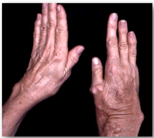
A sixty-three-year-old right hand dominant woman presented to our institution for evaluation and treatment of a complex deformity of her right upper extremity. Her examination showed multiple firm nodular soft-tissue masses on the dorsal and volar aspect of the right hand and wrist involving the thumb and index and long fingers. There were severe contractures of the index finger and first web space and small ulcerations with chronic infection in these areas. Dense sensory and motor neuropathy was present in the median nerve distribution in the right hand.
What was very unique in her medical history was a sequence of nodular mineralized masses that occurred from prior surgical procedures including the excision of her right little toe and later two surgical procedures to biopsy and then fill a large simple bone cyst in her right humerus with autogenous cancellous bone graft obtained from her right anterior iliac crest. Several years later she underwent a right carpal tunnel release with subsequent development on multiple nodules and contracture of the index finger.
An extensive laboratory evaluation including serum calcium, phosphorus, alkaline phosphatase, C-reactive protein and parathyroid hormone levels were all normal as was a 24-hour urine collection. An EMG revealed motor and sensory abnormalities of the axillary, radial, musculocutaneous , and median nerves.
Due to the severe functional compromise by the contracted index finger and first web space with chronic infection, we excised the second ray and a wide resection of the calcified masses over the volar and dorsal aspect of the right hand and wrist. A microneurolysis of the median nerve was done. The surgery was extremely tedious due to the compromised arterial circulation to the thumb. The index profundus tendon was routed around a pulley created with part of the flexor carpi ulnaris to restore thumb palmar abduction.
The question we considered preoperatively involved how to resurface the created defect given her propensity to develop these nodular masses at surgical sites. Given that all of her surgeries happened to involve the right hemicorporium, a decision was made to use a latissimus dorsi muscle flap harvested from the unaffected left side of the patient and revascularized with an end-to-side anastomosis to the ulnar artery in the mid forearm. A nonmeshed split-thickness skin graft was harvested from the contralateral buttock region and applied to the muscle.
At a five-year follow-up she regained protective sensation in the median nerve as well as improved side-to-side pinch. Noteworthy was the fact that no nodular masses occurred in the area of the latissimus flap as well as both of the contralateral donor areas.
Ultimately the condition was defined as focal fibronodular heterotopic ossification.
Figure 1: The appearance of the hand at presentation
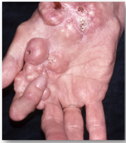
Figure 2: 5 years post-surgery with stable web without development of nodular masses in the web reconstruction